

Onderstaand artikel is geschreven in het Engels door oogarts Rick F. Sanchez
Following a cataract patient throughout the surgery day at AniCura SDU #
by Rick F Sanchez BSciBiol, DVM, DipECVO, CertVetEd / FHEA
EBVS Recognized European Specialist in Veterinary Ophthalmology
Cataract surgery removes the cloudy part of the lens in the animal’s eyes and replaces it with a lens implant to restore or improve vision. Cataracts can happen in young, middle aged and older animals alike, and they can be hereditary, be associated with diseases (i.e., diabetes in dogs and uveitis in cats), and they can also happen with age in dogs (i.e., age-related cataracts).
Cataract surgery has a good prognosis in small animals, and it has the chance to restore vision and dramatically improve the quality of life of an animal. Our ophthalmologist, Dr Rick F Sanchez, has a special interest in cataract surgery and has performed the operation close to a thousand eyes of dogs and cats over the last 20 years.
Before a cataract surgery
The process starts before the surgery day, with the first consultation with our ophthalmologist. The consult is used to decide if the patient is a good candidate for cataract surgery, and if unilateral or bilateral surgery is indicated for a particular surgical candidate.
The thorough eye exam and discussion with the owner takes approximately one hour and part of that time is reserved so Dr Sanchez can explain what cataracts, the possible risks of having cataracts and cataract surgery, the expected costs, the aftercare, and the re-examination schedule. Dr. Rick will also explain all the steps for the hospitalization and surgery.
What happens when your animal stays at AniCura SDU for cataract surgery? #
We know the families of the dogs and cats that stay with us for cataract removal cannot see every step of cataract surgery, and we would like to show you what the most important parts of this day look like for your beloved pet. The ten steps for hospitalization and surgery are listed below and include the following:
- Hospitalization (i.e., this is the process of admitting the patient into the hospital with all his / her medication, special food if needed, and a special toy or personal bed, though we also have our own at SDU)
- Ocular ultrasound (i.e., the first eye test on before the surgery (Figures 1 A and B))
- Preparing for the general anesthetic (Figure 2)
- ERG or electroretinogram (i.e., the second and last test before the surgery (Figures 3 A and B))
- Preparation of the operating room (i.e., this happens just before or during the ERG (Figures 4 A and B)
- Preparation of the eye/s for the surgery (Figure 5)
- Cataract surgery (Figures 6 A-D)
- Recovery and night stay (Figure 7)
- Eye exam the next day after surgery (i.e., the next morning, the surgeon re-examines the patient to make sure he / she is ready to go home).
- “Going back home!” (i.e., the surgeon and nursing team meet the owners, explain how things went and discuss the medications they need to give at home).
Surgery is teamwork
It is important to explain that cataract surgery requires a large team of people and interdisciplinary work (i.e., different specialists that work together to care for one patient). The ophthalmologist is the surgeon that examines the eye and performs the cataract removal surgery and places a new lens in the eye (i.e., a lens implant, Figure 6B) to restore vision.
A specialist in imaging performs the ocular ultrasound that looks inside the eye and measures the lens so the surgeon can find the right fit of lens implant (Figure 1B) and to rule out possible problems, such as a retinal detachment among other things. The specialist in anesthesia makes and adjusts the protocol for the general anesthesia needed for the surgery.
The nursing team cares for the patient every step of the way and is composed of the nurses in the front desk and the nurses of the surgery team that prepare the patient for anesthesia, help the surgeon prepare the operating room, monitor the patient during the operation, and care for the patient during the recovery period.
Cataract surgery for senior pets
Not all cataracts can undergo surgery, but most can. There is a myth that older animals should not have cataract surgery due to their age. However, age is not a disease, and older animals can undergo a safe anesthesia tailored to their needs to reduce preventable risks. Older animals that have stopped playing and sleep all day dramatically change their behavior after cataract surgery, when they can see again.
Cataract surgery can result in very happy pets when they can see again
Diabetic dogs often develop cataracts. They have additional risks that non-diabetic dogs do not have and should undergo cataract surgery without delay to avoid these, even if their sugar control is not completely stable yet.
We are dedicated to improving the lives of our companion animals
Dogs and cats that were previously blind, leave the hospital able to see their owners for the first time in weeks or even months, and there is nothing more gratifying for the surgeon and the nurses, and, of course, for the owners. If you have a patient with cataracts or have questions about the procedure or any of the steps it involves, please, do not hesitate to contact us at AniCura SDU.
Figure Legends
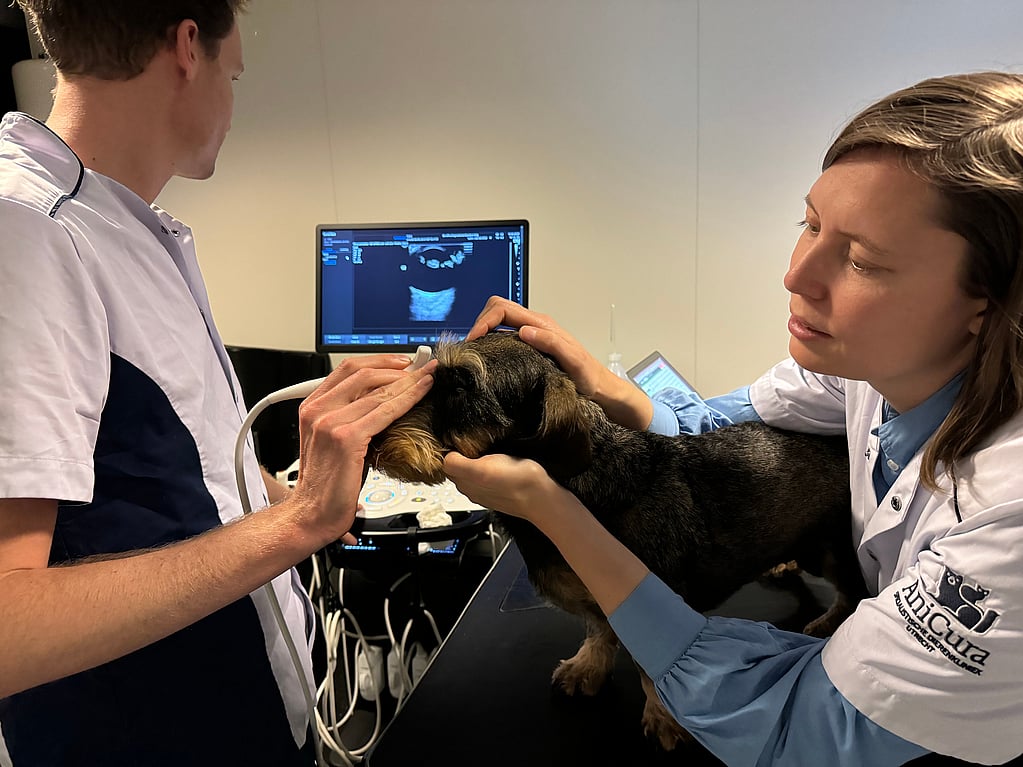

Figure 1. An ocular ultrasound is very well tolerated, and it is carried out with the cataract patient awake (A). A cataract is seen in the lens (i.e., the obvious white lines that make the ‘flying saucer’ shape inside the eye) and this is measured to give the surgeon an idea of what intraocular lens implant size to choose for the patient (B). It also serves to make sure there is no obvious disease behind the lens. At SDU we carry this test out on the day of surgery, as the results rarely leads to a cancellation of the surgery and we have the most up-to-date information on our patient.

Figure 2. The patient undergoing cataract surgery is anesthetized. The anesthesia team places an intravenous catheter as in this patient, and later administrates the drugs necessary for a safe anesthetic, intubates the patient to ensure we can bring oxygen and anesthetic gas to the patient, and breath for him / her, and we begin to monitor the patient vitals, which continues until after the surgery has finished and the patient has recovered.
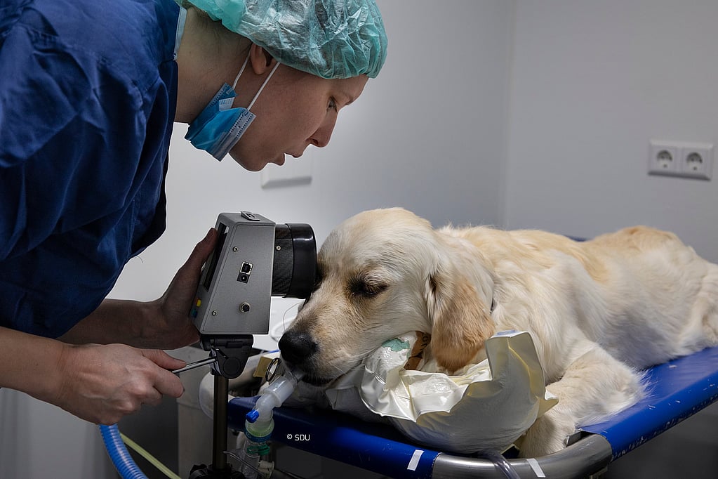
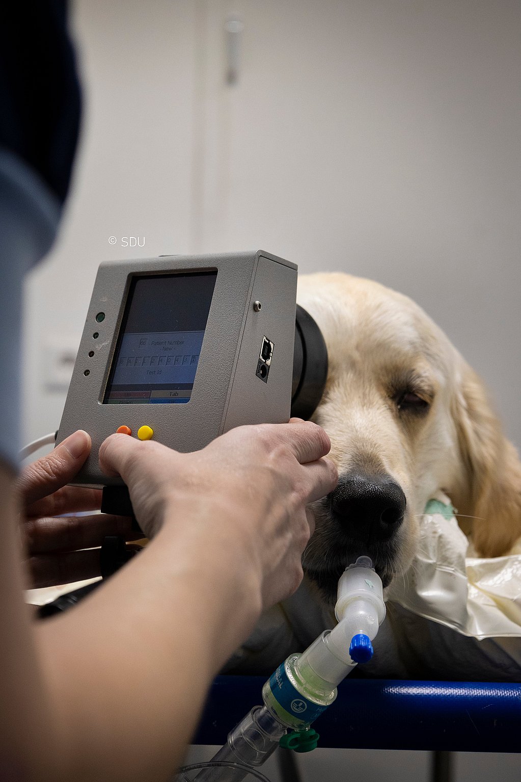
Figure 3. The patient undergoes a test called ERG (i.e., electroretinogram). It is similar to an EKG of the heart but instead of measuring the electrical activity of the heart, it measures the electrical activity of the retina in the eye, and, like the hear, it also produces a wave that we must interpret.


Figure 4. Preparing the operating room. The nurse and the surgeon prepare the multiple microsurgery instruments needed and place them in a specific order on the operating table (A). The surgeon also prepares the operating microscope (B) and the phacoemulsification machine (i.e., seen in image 6D as a screen with some numbers on it) that is used to remove the cataract.

Figure 5. Then, after the ERG and the preparation of the operating room, the nurse carefully aseptically prepares the patient’s eye/s (i.e., a special type of cleaning protocol) to make sure they are ready for the surgery.


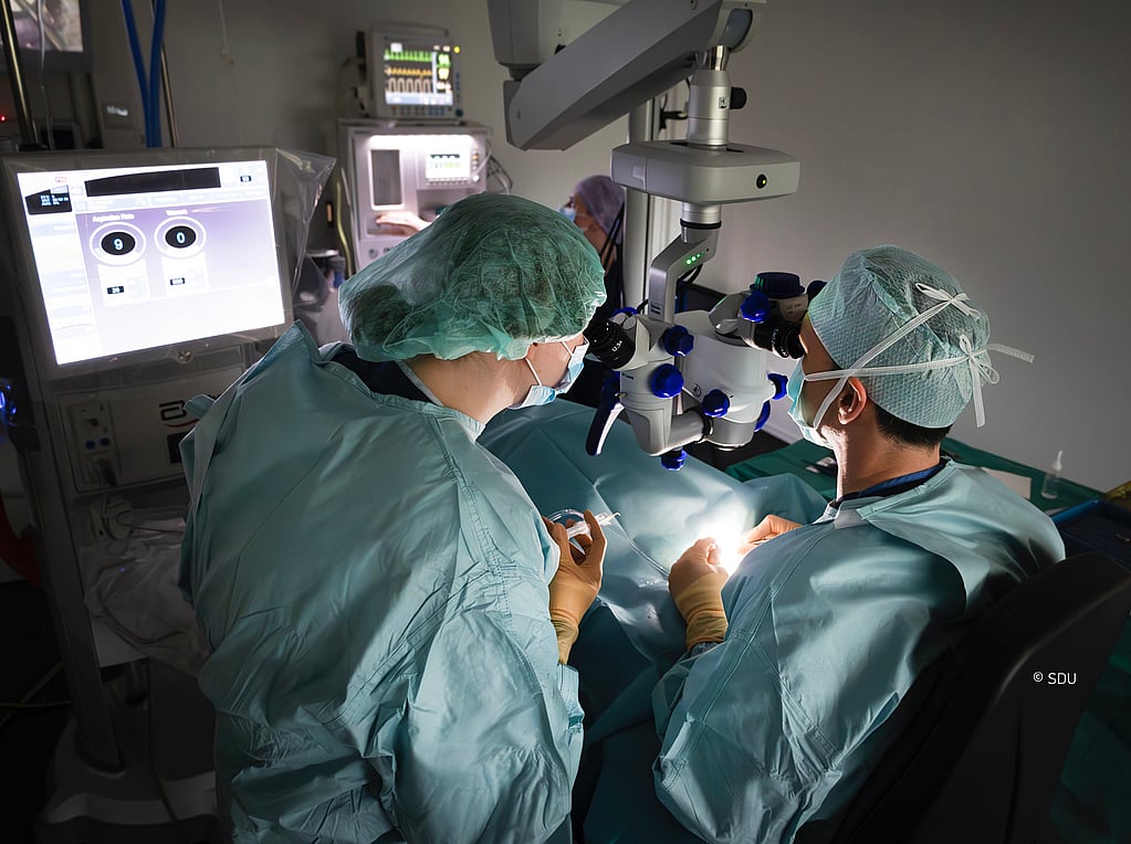
Figure 6. The surgeon makes tiny incisions in the periphery of the cornea (i.e., a 1mm and a 2.8 mm incisions) to have access to the cataract and remove it (A). Then, an intraocular lens implant is chosen (B) and is put into the eye (C), and after a few more steps to ensure the eye is ready, the small incisions are sutured closed (D).

Figure 7. Cataract surgery patients at SDU recover in a state-of-the-art ‘pod’ that has underfloor heating and glass doors where the nurses may write important information about the patient. The ‘pods’ also have special side access for intravenous fluids and additional warming and monitoring equipment. Cataract patients at SDU stay at the clinic one night with a dedicated night nurse that measures the eye pressure of the operated eye/s and makes sure the patient takes the medications he /she needs without the owners having to worry at home. There are separate rooms of ‘pods’ for dogs and cats.